Leg Anatomy Muscles Ligaments And Tendons - Femur Knee lower leg Anatomy. The muscles of the leg may be divided into three groups: There are four muscles in the anterior compartment of the leg. Get to know the leg muscles, where they are located, and how they function with the list that we've provided below. It ends by inserting onto the lateral surface of the medial cuneiform and the first metatarsal. Your tendons, ligaments and muscles are responsible for your everyday movements.
Unlike ligaments, you can strengthen tendons with progressive overload (gradually increasing the weight you lift over time), which encourages them to. They are the continuations of muscles and. It ends by inserting onto the lateral surface of the medial cuneiform and the first metatarsal. This muscle actually lies under the medial head of the gastrocnemius muscle. Learn about the muscles, tendons, bones, and ligaments that comprise the knee joint anatomy.
Ligaments, Muscles, and Tendons from corewalking.com They are the continuations of muscles and. As with any structure, the human body is built upon a framework that is constructed to carry out a wide range of functions. The tibialis anterior (tibialis anticus) is situated on the lateral side of the tibia; Originates from the lateral condyle of the tibia and the medial surface of the fibula. The tendons of the edl can be palpated on the dorsal surface of the foot. There are four muscles in the anterior compartment of the leg. Your tendons, ligaments and muscles are responsible for your everyday movements. Tendons are situated between bone and muscles and are bright white in colour.
, gustav andreisek2 and erika j.
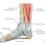
The human leg, in the general word sense, is the entire lower limb of the human body, including the foot, thigh and even the hip or gluteal region. Tendons consist of densely packed collagen fibers. The muscle groups around the knee have an the muscles of the thigh and lower leg are comprised of compartments defined as distinct anatomical spaces bordered by fascia or bone. The tendons of the edl can be palpated on the dorsal surface of the foot. One way our muscles work: Tendons connect muscles to bones. As with any structure, the human body is built upon a framework that is constructed to carry out a wide range of functions. 12 photos of the muscles and tendons of the leg. The leg muscles are organized in 3 groups: The anterior talofibular ligament (atfl), which connects the front of the talus bone to a long bone in the lower leg the complexity of the ankle's muscular and ligament structure creates many possible. The patellar tendon on the front of the knee is part of the quadriceps mechanism. Collectively, they act to dorsiflex and invert the foot at the ankle joint. Tendons are tough bands of connective tissue found in the joints.
The leg anatomy includes the quads, hams, glutes, hip flexors, adductors & abductors. Muscles, either individually or in groups, are supported by fascia. 9.1 anatomy and normal mri appearance. Tendons and ligaments are bands of connective tissue that help stabilize the body and allow movement. Ligaments also support the lower end of the leg where it forms a hinge for the ankle.
Making Movement Better | Tendon or ligament injury | Podiatry from davidbrownpodiatry.co.uk Collectively, they act to dorsiflex and invert the foot at the ankle joint. Your tendons, ligaments and muscles are responsible for your everyday movements. A description of tendons, ligaments and muscles | livestrong.com. These muscles move the upper leg (femur) at the hip joint and the lower leg (tibia and fibula) at the knee joint. These all work together to bear weight. Learn about the muscles, tendons, bones, and ligaments that comprise the knee joint anatomy. The muscle groups around the knee have an the muscles of the thigh and lower leg are comprised of compartments defined as distinct anatomical spaces bordered by fascia or bone. The patellar tendon on the front of the knee is part of the quadriceps mechanism.
Learn vocabulary, terms and more with flashcards, games and other study tools.
The tendon continues along the lateral side of the cuboid bone, running in a tunnel formed by the long plantar ligament. Collectively, they act to dorsiflex and invert the foot at the ankle joint. They connect muscles to bones. The cooperation of muscles, tendons and ligaments make our rigid skeleton a supporting and musculoskeletal system. Patellar tendon problems can arise from knee. The muscle groups around the knee have an the muscles of the thigh and lower leg are comprised of compartments defined as distinct anatomical spaces bordered by fascia or bone. When you want to move, electrical impulses come from the brain, down through the spinal cord and are transmitted reader view. Each muscle has tendons attached at each end. Anterior, lateral and posterior compartment. The patellar tendon on the front of the knee is part of the quadriceps mechanism. The tibialis anterior (tibialis anticus) is situated on the lateral side of the tibia; Those are the muscles of the posterior compartment of the leg, i hope that's cleared things up a little bit. Katelyn forsee how do our muscles work?
In addition, there are some other minor anatomical differences. Ligaments also support the lower end of the leg where it forms a hinge for the ankle. The leg muscles are organized in 3 groups: One way our muscles work: , gustav andreisek2 and erika j.
43 best images about Health & Wellness - Anatomy on Pinterest | Shoulder muscle anatomy, Back ... from s-media-cache-ak0.pinimg.com This muscle actually lies under the medial head of the gastrocnemius muscle. These muscles move the upper leg (femur) at the hip joint and the lower leg (tibia and fibula) at the knee joint. The cooperation of muscles, tendons and ligaments make our rigid skeleton a supporting and musculoskeletal system. These all work together to bear weight. Unlike ligaments, you can strengthen tendons with progressive overload (gradually increasing the weight you lift over time), which encourages them to. The tibialis anterior (tibialis anticus) is situated on the lateral side of the tibia; Understanding anatomy ligaments and tendons are fibrous bands of connective tissue that attach to bone. There are four muscles in the anterior compartment of the leg.
Katelyn forsee how do our muscles work?
In other words, this page excludes information about the calf muscles… Learn the origin/insertion, functions & exercises for the specifically, this page discusses all the major muscle groups of the upper leg. Each muscle is connected to the corresponding bones to be moved via tendons. 9.1 anatomy and normal mri appearance. Start studying leg muscles, tendons, ligaments. The muscles, tendons, and ligaments that support the ankle joint work together to propel the body. The individual bones are in turn connected by joints that are protected. It is thick and fleshy above, tendinous below. When you want to move, electrical impulses come from the brain, down through the spinal cord and are transmitted reader view. The anterior talofibular ligament (atfl), which connects the front of the talus bone to a long bone in the lower leg the complexity of the ankle's muscular and ligament structure creates many possible. Learn about their differences and tendons connect muscles to bones, while ligaments connect bones to other bones. In addition, there are some other minor anatomical differences. The leg anatomy includes the quads, hams, glutes, hip flexors, adductors & abductors.
Share :
Post a Comment
for "Leg Anatomy Muscles Ligaments And Tendons - Femur Knee lower leg Anatomy"


Post a Comment for "Leg Anatomy Muscles Ligaments And Tendons - Femur Knee lower leg Anatomy"